|                                                                ISSN: 2997-321X                                               Doi: 10.47991/2997-321X/AJCRR-114  

A Diagnostically Challenging Case Post Induction of General Anaesthesia for Endovascular Aneurysm Repair: Anaphylaxis, Takotsubo, Kounis Syndrome or ATAK, A Case-Report Based Mini Review

Author(s): Papageorgiou Lampriani¹*, Christoforou Panagitsa², Kounnos Christos², Bekos Christos², Kiriakou Maria¹
¹Department of Anaesthesiology, General Hospital of Nicosia, Nicosia, Cyprus
²Department of Vascular Surgery, General Hospital of Nicosia, Nicosia, Cyprus
Papageorgiou Lampriani*
Address: Thessalonikis 9 street, Ormidhia, 7530, Larnaca, Cyprus. E-mail: lambrie_pap@hotmail.com; Tel: 00357-22604401/ 00357-99599185
Citation: Papageorgiou L, Christoforou P, Kounnos C, Bekos C, Kiriakou M (2024) A Diagnostically Challenging Case Post Induction of General Anaesthesia for Endovascular Aneurysm Repair: Anaphylaxis, Takotsubo, Kounis Syndrome or ATAK, A Case-Report Based Mini Review. American J Cas Rep Rev: AJCRR-114. 
Received: 07 November, 2024
Accepted: 14 November, 2024
Published: 20 November, 2024
Abstract
Takotsubo cardiomyopathy or stress-induced cardiomyopathy is an apical balloon like syndrome of left ventricle during systole with absent coronary obstructive disease on angiogram. This syndrome was named after an “octopus-catcher” used in Japan called “takotsubo” discovered in the late 80s. This so called “broken heart syndrome” is the result of extreme physical, emotional and psychological stressors. Iatrogenic causes can also promote Takotsubo cardiomyopathy, enhancing the theory of catecholamine toxicity as the main pathogenetic cause behind this syndrome. Sympathomimetic drugs can cause this not so rare disease nowadays with adrenaline being the most well-known causative agent. Frequently, adrenaline is administered as a first line treatment consisting a cornerstone therapy of anaphylaxis and all kinds of allergic and anaphylactoid reactions. It is not rare for adrenaline to cause takotsubo cardiomyopathy leading to catecholamine toxicity in the body, especially when administered intravenously. Kounis syndrome is a type of acute coronary event caused by inflammatory mediators which are released in some allergic reactions causing coronary vasospasm. ATAK complex is a combination of Adrenaline, Takotsubo, Anaphylaxis and Kounis syndrome which was very recently reported in literature. It was only a few years ago when it was discovered that there seems to be an interesting connection between these different clinical entities with common pathogenetic and pathophysiological pathways. In this review, we present a case with Takotsubo cardiomyopathy and possibly anaphylaxis and ATAK complex who arrested immediately post general anaesthesia induction for endovascular aneurysm repair of left common iliac artery and abdominal aorta aneurysms.
Keywords: adrenaline induced cardiomyopathy, Takotsubo cardiomyopathy, drug-induced cardiomyopathy, iatrogenic Takotsubo cardiomyopathy, catecholamine toxicity, Kounis syndrome, ATAK (Adrenaline, Takotsubo, Anaphylaxis and Kounis syndrome).
Introduction
Takotsubo cardiomyopathy (TCM) is a reversible cardiac syndrome associated with transient left ventricular dysfunction of the apical and mid segments of the left ventricle. It occurs in 0.7%-2.5% of patients presenting with symptoms of acute coronary syndrome. [1] TCM is characterised by a unique morphological feature of apical ballooning of the left ventricle during systole. It occurs as a result of intense emotional and physical stress in the absence of coronary artery disease and myocarditis. It is also known as stress cardiomyopathy or “broken heart syndrome”. Catecholamine toxicity is known to play vital importance in the pathogenesis and pathophysiology of the syndrome, therefore not only emotional stress can induce its phenotype, rather a catecholamine surge overload caused by either physical, emotional and even iatrogenic causes [2, 3].
The diagnosis is set in the presence of the following criteria: regional wall motion abnormalities extending beyond the territories of an epicardial vessel, unobstructed coronary arteries, elevated troponin and/or new electrocardiographic ischemia findings and the absence of active myocarditis and pheochromocytoma [4]. The history of the disease lies back in 1983, where Dove et al. reported a case of a patient who presented to a Japanese hospital with acute myocardial infarction [5]. The image of the patient’s left heart resembled an octopus trapping pot (a “takotsubo”) which has a round bottom and a narrow neck, where the cardiomyopathy took its name from [6]. Multiple similar cases from Japan followed this report whereas worldwide cases were described later [7-10]. The initial cause of this syndrome was thought to be multivessel epicardial spasm resulting in myocardial stunning [11].
Many subtypes have been described since its discovery, but two main types exist: typical TCM which involves apical hypokinesia/akinesia segments and the atypical form which is actually its reverse type, meaning basal or mid wall hypokinesis/akinesis segments and/or even right ventricular ballooning [4]. The most common presenting symptoms are angina type chest pain and dyspnoea [12]. Cardiac arrest, syncope and arrythmias can also be present [13]. In critically ill patients, symptoms may include worsening of their clinical condition. The syndrome can be incidentally discovered, either by electrocardiographic findings, echocardiography, increased myocardial biomarkers and coronary angiogram [14]. Signs and symptoms are non-specific and are similar to those with acute myocardial infarction and/or acute heart failure, therefore do not help in the differential diagnosis.
The mechanisms of the disease have yet to be elucidated. It is believed that coronary artery vasospasm, microcircular dysfunction and transient obstruction of the left ventricle outflow tract (LVOT) play a crucial role in the pathogenesis of the TCM. Catecholamine overload serves as the cornerstone and initial cause of a domino facts leading to the disease. This cascade can ultimately lead to substantial alterations of myocardial structure including increased extracellular matrix, contraction band necrosis and neutrophil infiltration. Contractile dysfunction can be the result of increased oxidative stress and the alteration of Ca2+ handling proteins [1].
This direct release of catecholamines into the myocytes decrease myocardial viability through cAMP mediated calcium overload through activation of adrenergic receptors resulting in myocardial toxicity. It has been proposed that the activation of Gs type receptors can switch into type Gi, therefore leading to the opposite effect, meaning decreased contractility. This negative inotropic effect might explain one of the protective mechanisms which can explain why this syndrome is temporary since most patients regain full function of their heart with no compromised effect in a short period of time, possibly two to four weeks. Another reasonable theory explaining the differences of myocardial segments in contractility is the fact that B2 adrenoreceptors are mainly located in the apex rather than the basal segment of the left ventricle, whereas B1 are more expressed in the base and mid segments rather than the apical region of the heart. B2 adrenergic stimulation is of Gi type, therefore reduced sympathetic tone on the heart, whereas B1 is of Gs, meaning an enhanced sympathetically driven inotropic effect [15].
There is no specific treatment of TCM, therefore the treatment is only supportive as this process is transient and, in most cases, reversible. Mortality rate is similar to the mortality rate of an acute ST elevation infarction, around 4-5%. Despite substantial improvement in understanding of the syndrome, a number of significant gaps of knowledge still remains [15].

Kounis syndrome or Kounis hypersensitivity-associated coronary syndrome is a type of acute coronary syndrome as a result of coronary vasospasm which is induced by an acute anaphylactic reaction to a trigger. It was first reported by Kounis and Zavras in 1991 [16]. In more recent years, a connection between Takotsubo cardiomyopathy and Kounis syndrome has been made since there are many similarities between these entities and common pathogenetic and pathophysiological mechanisms. The ATAK complex (Adrenaline, Takotsubo, Anaphylaxis and Kounis syndrome) is the concurrent incidence of adrenaline administration as a result of anaphylaxis, the presence of Takotsubo cardiomyopathy, anaphylaxis and Kounis hypersensitivity-associated coronary syndrome. A few cases have been reported worldwide in various triggers [17-21].

In this article we report the case of a patient who presented with Takotsubo cardiomyopathy after administration of a single bolus dose of 1 mg adrenaline after he arrested a few seconds post induction of general anaesthesia (GA) which initially was treated as anaphylactic shock possibly related to rocuronium administration for endovascular aortic repair (EVAR) surgery. Kounis syndrome could not be excluded in this case.
Case report
We present the case of a 75-year-old Caucasian man who was admitted to the hospital for endovascular repair of his left common iliac saccular aneurysm of 19×22 mm in diameter and 27 mm in length and a bulge of the abdominal aorta graft repair. This patient suffered from hypertension, dyslipidaemia, coronary artery disease ((percutaneous coronary insertion (PCI) to left anterior descending (LAD) artery in 2022 with ejection fraction (EF) 50-55%)), peripheral vascular disease and an undiagnosed spine tumour between T12-L5 which was probably haemangioma and was an ex-smoker. He was otherwise fit and healthy with good performance status. He had previously been under anaesthesia for multiple surgeries with no noted problems. Preoperative electrocardiogram (ECG), chest X-ray (CXR) and transthoracic echo (TTE) were unremarkable.

Anaesthesia started with the insertion of two peripheral lines on the left hand and the insertion of a left radial artery. Monitoring was performed with continuous ECG, invasive blood pressure (IBP), non-invasive blood pressure (NIBP), capnography, bi-spectral index (BIS) values (entropy) and oximetry. The patient was given some volume of intravenous fluids and was pre-oxygenated for at least five minutes prior to induction. Preoperative vital signs were normal. GA was started with the administration of fentanyl, propofol and rocuronium while 100 μgrs of phenylephrine was given bolus a few seconds later for mildly low blood pressure (BP). The patient was manually ventilated for two minutes post the administration of rocuronium and was intubated with an endotracheal 8.5 mm tube with no difficulties. Seconds after intubation, the patient abruptly became extremely hypotensive ((systolic blood pressure (SBP)=60 mmHg)) and tachycardic ((heart rate (HR)=120-130 beats per minute (bpm)) and no breathing sounds could be heard whereas capnography showed the presence of exhaled carbon dioxide and an obstructive airway pattern with normal airway pressures. No rush or wheezing or airway oedema were present at the time. He was quickly given adrenaline bolus of 50 μgr bolus intravenously as acute anaphylaxis was suspected but no immediate effect was seen. He was administered another 200 μgr of adrenaline bolus intravenously but again no direct response was seen.
Furthermore, the patient was started additional boluses of intravenous fluids at the time of acute hypotension. After a few seconds, his invasive systolic blood pressure fell to 30 mmHg. At this point with no extra delays, he was immediately given the rest adrenaline dose of 750 μgr bolus intravenously as the patient was in cardiac arrest (pulseless electrical activity) and chest compressions were commenced. Hydrocortisone and an antihistamine bolus were also administered intravenously while he continued to have boluses of crystalloids. The patient’s response to GA induction was treated at first as severe anaphylactic shock possibly related to rocuronium administration with the administration of a small dose of intravenous adrenaline and subsequently treated as a cardiac arrest with the administration of a total full dose of 1 mg intravenous adrenaline as per the cardiac arrest protocol.

Seconds after the administration of bolus adrenaline, the patient became extremely hypertensive (systolic BP=260-280 mmHg) and tachycardic and presented a lot of arrythmias on the monitor including extra-systoles and ventricular tachycardia of around 180 bpm while remained extremely hypertensive. Boluses of nitroglycerin were then administered intravenously, and the patient’s blood pressure started to gradually decrease, to a point where he actually needed again boluses of vasopressors (systolic BP=50 mmHg). Noradrenaline infusion was started and titrated to efficacy, while boluses of phenylephrine were also occasionally administered. The patient’s blood pressure started again to rise and so noradrenaline titrated downwards so as for blood pressure to stabilise to normal values. Indeed, the patient’s blood pressure was stabilised approximately 20-30 minutes post the acute hypotension episode. No additional bolus of adrenaline was administered.
 
After stabilisation of the patient’s condition, we proceeded to the angiogram to exclude any aneurysm rupture because of the very high values of blood pressure we had previously seen on the monitor. After puncturing the right common femoral artery, an angiographic examination and digital subtraction angiography (DSA) were performed immediately, with no rupture found, therefore we decided to proceed further with the procedure of endovascular repair of his aneurysms. After an open preparation of the left femoral artery, embolization of the left internal iliac artery was initially performed using ruby coils and then an AFX2 BEA22-70/I16-30 and left iliac extension of the Endurant Medtronic stent graft system ETLW161093EE were deployed. The final angiographic result was satisfactory, without any thrombosis or endoleak present, with complete exclusion of the aneurysms (Fig. 1). During endovascular therapy, the patient was stable, with a gradual reduction of vasopressor drugs. However, due to the previous episodes of hemodynamic instability, it was deemed necessary to transfer the patient to Intensive Care Unit (ICU).
Figure 1: DSA depicting endovascular repair of the left iliac artery and abdominal aorta aneurysms. After an open preparation of the left femoral artery, embolization of the left internal iliac artery using ruby coils and then an AFX2 BEA22-70/I16-30 and left iliac extension of the Endurant Medtronic stent graft system ETLW161093EE were deployed. The final angiographic result revealed no extravasation, thrombosis or endoleak, with complete exclusion of the aneurysms.
DSA: Digital Subtraction Angiography
The operation lasted approximately three hours, the patient was administered three litres of intravenous crystalloids and urinated around one and a half litres. No blood products were given while no additional neuromuscular blockers were administered. He was sedated with propofol and noradrenaline infusion was titrated to blood pressure normal values. Arterial blood gas values were unremarkable (lactate was eventually reduced to normal values from a maximum of 3.5 mmol/l).

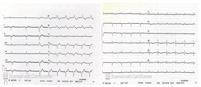
During hospitalisation to ICU, a few hours later after the transfer, the patient gradually became hypotensive and required increased dosage of noradrenaline for normalisation of his blood pressure. He had a bedside echocardiography which showed reduced EF around 30% and regional wall motion abnormalities with apical hypokinesia. ECG showed new T wave inversion in anterolateral leads (fig.2) and troponin was positive and continued to rise on the second measurement. On the same day, he was led to catheterization laboratory for coronary angiogram after careful consideration of all related risks and benefits.
Figure 2: ECGs on admission to ICU: 1st ECG on admission to ICU showing alternate SR and bradycardia of around 35bpm with junctional rhythm and RBBB. 2nd ECG showing SR with TWI on lateral and anterior leads implying acute ischemia to the anterior and lateral walls of the LV.
bpm: beats per minute, ECG: electrocardiogram, ICU: Intensive Care Unit, LV: left ventricle, RBBB: right bundle branch block, SR: sinus rhythm, TWI: T – wave inversion.
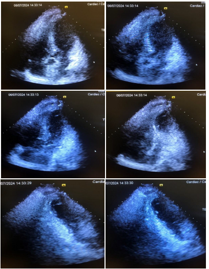
He remained sedated and intubated up to that time. Coronary angiogram showed non-significant coronary artery disease (fig.3) and severe cardiogenic shock was indeed verified by invasive cardiologists with an EF of left ventricle (LV) around 20% and akinesia of apical segments and LVOT obstruction resembling typical Takotsubo cardiomyopathy (fig.4 and 5). The administration of adrenaline for cardiac arrest as a cause of suspected acute anaphylactic reaction possibly to rocuronium administration, troponin rise, new ECG ischemia findings, transient severe systolic impairment of the LV and the morphological feature of ballooning of the LV with unobstructed coronary arteries is consistent with the diagnosis of possibly iatrogenic adrenaline induced Takotsubo cardiomyopathy.
Figure 3: Coronary angiogram of the patient with non-significant coronary artery disease. Coronary angiogram revealed no significant obstructed coronary arteries.
Figure 4: Aortogram of patient exhibiting Takotsubo cardiomyopathy features. Aortogram showing apical ballooning during systole with apical hypokinesia and good contractility of only basal and mid-segments of the patient’s heart.
Figure 5: Echocardiography of patient showing Takotsubo cardiomyopathy. Echocardiography at the catheterisation laboratory showing an apical balloon like cardiomyopathy, setting the diagnosis of Takotsubo cardiomyopathy.
The tryptase levels post arrest at t=0 hrs and t=3 hrs post the event after an allergology assessment both came back negative. It is important to mention here again that the patient had a normal echocardiography six months ago.

Surprisingly, our patient was extubated two days later after the event. He claimed he felt good at first, although a few hours later, he developed acute back pain and morphine was administered with some remission of his pain. ECG remained the same and troponin was reduced from previous measurements. Arterial blood gas though showed increase of lactate to 4 mmol/l despite adequate intravenous filling. An abdominal aorta and iliac arteries computed tomography angiography (CTA) was immediately performed with no signs of haemorrhage, endoleak or thrombosis present. At night, the patient became extremely tachycardic presenting fast atrial fibrillation (up to 170 bpm) while hemodynamically unstable. He received synchronised cardioversion twice with restoration back to normal sinus rhythm. He was a little confused post the event but able to obey commands and move all limbs normally. He continued to improve the following days and subsequently was discharged from ICU six days later. Echocardiography three days later showed akinetic apical and medial segments with satisfactory kinesis of basal segments, an EF around 30-35%, mild mitral regurgitation (MR) with no systolic anterior motion (SAM) phenomena, a moderately dilated right ventricle (RV) with preserved systolic function, moderate tricuspid regurgitation (TR) and right ventricular systolic pressure (RVSP) of 45-50 mmHg. The patient received antiplatelet treatment with aspirin upon discharge, however, after a cardiological evaluation, he received a new oral anticoagulation with Dabigatran due to an episode of atrial fibrillation during his hospitalisation to the ICU.
A few days post discharge from ICU, the patient developed bilateral pleural effusions and was assessed by cardiology who requested a computed tomography pulmonary angiogram (CTPA) which confirmed the previous diagnosis and excluded any large pulmonary thrombosis. Paracentesis of the pleural effusions showed to be transudate. Echo revealed dilatation of the right chambers, severe TR, RVSP of approximately 60 mmHg and no signs of pericardial effusion. Moreover, the patient’s EF improved to preoperative levels. Diagnosis of acute right heart failure was set and increased diuresis followed, which ultimately led to improvement of the patient’s symptoms. The patient continued to improve and was discharged from the hospital a few days later with follow up with vascular surgery and cardiology. Two weeks exactly post the arrest, he was assessed by his cardiologist who performed another echo which showed normal LV size, wall thickness and EF, mild dilatation of the ascending aorta (up to 42.2 mm) with normal aortic root, impaired relaxation of LV, mild dilatation of RV with preserved function, mild MR and moderate TR and mildly elevated pulmonary artery pressure ((estimated mean pulmonary artery pressure (mPAP)=25 mmHg)).
Discussion
The possibility of Takotsubo cardiomyopathy prior to induction of GA presenting as an arrest is also possible in this scenario but the patient did not mention any dyspnoea symptoms before to his past medical history. Nevertheless, he had reasons of Takotsubo cardiomyopathy as he had recently lost his wife. The case of anaphylaxis could not be excluded in this case but could not be verified either. It is important to remember that skin reactions in anaphylactic shock are absent in about 20% of the people [22]. In addition, tryptase levels can be negative in a significant number of anaphylaxis cases [23]. Acute serum tryptase level carries high positive predictive value and specificity but poor sensitivity and negative predictive value [24]. It is important to have these facts in mind because in our case, anaphylaxis could not be excluded as the initial cause of perioperative shock, although eventually his shock became cardiogenic in origin. Antibiotics and muscle relaxants have been well accused as allergic triggers between all medication [25]. We assume that our patient collapsed right after induction of GA as a result of sensitization to rocuronium from previous surgeries presenting with cardiac arrest a few seconds post intubation. The 1 mg of adrenaline which was administered intravenously led to a catecholamine explosion which ultimately caused him Takotsubo cardiomyopathy as it was later confirmed by coronary angiogram and echocardiography. Of course, Kounis syndrome could not be excluded either in this case. It was certainly a very diagnostically challenging case since any diagnosis could only be verified or excluded retrospectively. ATAK complex should also be involved in differential diagnosis, although a very few cases have been published worldwide reporting this complex quadrable entity as the primary diagnosis caused by various triggers, drugs or not [17-21, 26].
We searched the literature in Pubmed performing advanced search using the terms “takotsubo” AND “adrenaline administration” or “adrenaline induced” or “iatrogenic” to be present in title or abstract and we found 59 results. On a second attempt, we searched the literature in Pubmed using the terms “takotsubo” AND “adrenaline-induced” or “iatrogenic” and/or “kounis” or “ATAK complex” and we have found 45 articles. Of these 104 articles, we included 48 articles (33 and 15 respectively from the two previous search attempts respectively) as relevant to the subject of question as they did not meet any exclusion criteria of which 3 articles were in double. Therefore, 45 articles were included for further research in this case-based review.

There are several reports about iatrogenic Takotsubo cardiomyopathy confirming the hypothesis that this syndrome is the result of extreme catecholaminergic stress caused in the body not only because of physical, emotional and psychological stressors but also due to iatrogenic causes, mainly sympathomimetic drugs [27-35]. Diagnostically, TCM always seems to be a challenging case [29, 36]. Interestingly, catecholamine toxicity seems to be implicated in the pathogenesis of “broken heart syndrome” [36-40]. The last list of possible drug triggers of TCM included 72 drugs and was last updated in 2022 [41, 42]. Another recent review provided additional evidence about a sum of 75 drugs. [43] The majority generate sympathetic overstimulation. Nevertheless, some of the drugs mentioned have no sympathomimetic activity [41, 43].
Adrenaline is however the most known and well-discussed drug which has been accused of inducing Takotsubo cardiomyopathy to patients [3, 35, 37, 40, 44-53] – even atypical forms of the disease – and possibly noradrenaline is the second [35, 54-56]. Undoubtedly, it requires extreme caution when adrenaline is to be given intravenously and adequate heart monitoring and cardiology skills are essential in these cases due to myocardial stunning [46].

Anaphylaxis itself might play a role in the pathogenesis of TCM while adrenaline to treat it might mask the effects of anaphylaxis to this clinical syndrome. Echocardiography skills at the bedside from the very beginning of the patient’s assessment could help to delineate the facts in this complex and complicated sequence of events following acute shock, any type of that might be [57]. It is interesting to mention here that myocardial injury as a result of anaphylaxis can been seen in up to 7-8% of cases [58].

Kounis syndrome is the concurrence of acute coronary syndromes as the result of epicardial coronary vasospasm with conditions associated with mast cell activation such as allergies or hypersensitivity or anaphylactic or anaphylactoid reactions [59]. ATAK complex is the association of Adrenaline, Takotsubo, Anaphylaxis and Kounis syndrome as already mentioned. Kounis syndrome and takotsubo present many similarities and common pathogenetic and pathophysiological mechanisms. These syndromes are frequently bound and masked by adrenaline administration, especially when anaphylaxis is suspected. Regardless of that, adrenaline mediated stress, either endogenous or exogenous, could be the link between these two clinical entities. Distinction between these two syndromes is often difficult and challenging or even unnecessary since treatment is commonly supportive [17, 60-62]. Nevertheless, a very few reports in literature have been documented as ATAK complex cases since this clinical combination came on the surface a very few years ago [18-21, 26, 61]. Of course, there are cases documented in literature which fall under this category, but they were not labelled as such [63, 64].
Kounis et al. speculated some of the basic pathophysiological mechanisms behind Kounis syndrome and gave emphasis on the inflammatory mediators which are released by mast-cells, eosinophils and other cells which are involved in the pathogenesis of inflammation as a final trigger pathway leading to vasospasm, the formation of thrombi or rupture of a culprit plaque in a vessel. Those mediators include proteases such as tryptase and chymase, arachidonic acid products, histamine, platelet activating factor and a variety of cytokines and chemokines released in the activation process. Kounis syndrome can frequently complicate anaesthesia, vaccination, medical therapy and stent implantation and can be associated with other vasculopathies and takotsubo syndrome. Nevertheless, some of the inflammatory mediators and mechanisms mentioned above seem to be common with events of non-allergic aetiology [59]. Delineation of those molecular mechanisms is of great importance as to gain better understanding of those syndromes and address targets for molecular therapy in the future.
The same pathophysiology of myocardial injury seems to be common between Kounis syndrome and Covid-19 infection. Sars-cov2 invades the cells through angiotensin converting enzyme 2 (ACE2) receptors located in different types of human cells. This leads to downregulation of the ACE2 receptors resulting in prothrombotic events such as the haemostatic imbalance via activation of the coagulation cascade, impaired fibrinolysis, thrombin generation, vasoconstriction, endothelial and platelet activation and pro-inflammatory cytokine release. The ACE2, renin-angiotensin-aldosterone system (RAAS) and kinin-kallikrein system (KKS) are the main proposed mechanisms contributing to cardiovascular complications associated with Covid-19 infection. The same mechanisms might be implicated in cardiovascular morbidity associated with Kounis syndrome, Takotsubo cardiomyopathy, heart failure, hypertension, myocarditis, microvascular disease, cardiac arrythmias, strokes and ischemic myocardial injuries, as well as prolonged Covid [65].
ATAK complex is underdiagnosed among physicians worldwide. It is important to shed light to the common and non-common pathways of the constituents of this clinical quadrable combination. If we understand it better, we will be able to recognise and treat it better as physicians early. It is not uncommon for all diagnoses of this complex to be set retrospectively and separately. Therefore, better knowledge of these syndromes and pathogenetic mechanisms as well as awareness of their interconnection is of vital importance between doctors since early suspicion and treatment not only can save patients’ lives but can lead to better human care and less iatrogenic complications. Hopefully, this knowledge can ultimately lead to the discovery of specific molecular target therapies and better understanding of the pathophysiology and pathogenesis of so different, yet partially common causative pathways of a plethora of cardiovascular diseases.
Additional materials: No.
Acknowledgements: Not applicable.
Funding: Not applicable.
Availability of supporting data: Not applicable.
Ethical approval and consent to participate: No IRB approval required.
Competing interests: The authors declare that they have no competing interests.
References
1. Nef, H.M., et al., Mechanisms of stress (Takotsubo) cardiomyopathy. Nature Reviews Cardiology, 2010. 7(4): p. 187-193.
2. Amin, H.Z., L.Z. Amin, and A. Pradipta, Takotsubo Cardiomyopathy: A Brief Review. J Med Life, 2020. 13(1): p. 3-7.
3. Nazir, S., et al., Takotsubo cardiomyopathy associated with epinephrine use: A systematic review and meta-analysis. Int J Cardiol, 2017. 229: p. 67-70.
4. Scantlebury, D.C. and A. Prasad, Diagnosis of Takotsubo cardiomyopathy. Circ J, 2014. 78(9): p. 2129-39.
5. Aizawa, K. and T. Suzuki, Takotsubo cardiomyopathy: Japanese perspective. Heart Fail Clin, 2013. 9(2): p. 243-7, x.
6. Sato, H., Tako-tsubo-like left ventricular dysfunction due to multivessel coronary spasm. Clinical aspects of myocardial injury : From ischemia to heart failure, 1990: p. 56-64.
7. Desmet, W.J., B.F. Adriaenssens, and J.A. Dens, Apical ballooning of the left ventricle: first series in white patients. Heart, 2003. 89(9): p. 1027-31.
8. Bybee, K.A., et al., Clinical characteristics and thrombolysis in myocardial infarction frame counts in women with transient left ventricular apical ballooning syndrome. Am J Cardiol, 2004. 94(3): p. 343-6.
9. Pavin, D., H. Le Breton, and C. Daubert, Human stress cardiomyopathy mimicking acute myocardial syndrome. Heart, 1997. 78(5): p. 509-11.
10. Nascimento, F.O., et al., The characteristics of stress cardiomyopathy in an ethnically heterogeneous population. Clinics (Sao Paulo), 2011. 66(11): p. 1895-9.
11. Dote, K., et al., [Myocardial stunning due to simultaneous multivessel coronary spasms: a review of 5 cases]. J Cardiol, 1991. 21(2): p. 203-14.
12. Tsuchihashi, K., et al., Transient left ventricular apical ballooning without coronary artery stenosis: a novel heart syndrome mimicking acute myocardial infarction. Angina Pectoris-Myocardial Infarction Investigations in Japan. J Am Coll Cardiol, 2001. 38(1): p. 11-8.
13. Pant, S., et al., Burden of arrhythmias in patients with Takotsubo cardiomyopathy (apical ballooning syndrome). Int J Cardiol, 2013. 170(1): p. 64-8.
14. Chockalingam, A., et al., Acute left ventricular dysfunction in the critically ill. Chest, 2010. 138(1): p. 198-207.
15. Pelliccia, F., et al., Pathophysiology of Takotsubo Syndrome. Circulation, 2017. 135(24): p. 2426-2441.
16. Kounis, N.G. and G.M. Zavras, Histamine-induced coronary artery spasm: the concept of allergic angina. Br J Clin Pract, 1991. 45(2): p. 121-8.
17. Ballesteros, R.V., et al., Kounis and Takotsubo, Two Syndromes Bound by Adrenaline: The “ATAK” Complex. Case Rep Cardiol, 2023. 2023: p. 7706104.
18. Kounis, N.G., et al., The ATAK complex (Adrenaline, Takotsubo, Anaphylaxis, and Kounis hypersensitivity -associated coronary syndrome) in neurological conditions. Indian J Crit Care Med, 2016. 20(4): p. 255-6.
19. Margonato, D., et al., Takotsubo cardiomyopathy associated with Kounis syndrome: A clinical case of the “ATAK complex”, in J Cardiol Cases. 2019: Japan. p. 52-56.
20. Minciullo, P.L., et al., ATAK Complex (Adrenaline, Takotsubo, Anaphylaxis, and Kounis Hypersensitivity-Associated Coronary Syndrome) after COVID-19 Vaccination and Review of the Literature, in Vaccines (Basel). 2023: Switzerland.
21. Alarcón Gallardo, E., R. Escudero Apesteguía, and C. Sanz Bescós, ATAK Complex due to Amoxicillin: A Case Report. J Investig Allergol Clin Immunol, 2024. 34(1): p. 62-64.
22. Brown, S.G., R.J. Mullins, and M.S. Gold, Anaphylaxis: diagnosis and management. Med J Aust, 2006. 185(5): p. 283-9.
23. Caughey, G.H., Tryptase genetics and anaphylaxis. J Allergy Clin Immunol, 2006. 117(6): p. 1411-4.
24. Buka, R.J., et al., Anaphylaxis and Clinical Utility of Real-World Measurement of Acute Serum Tryptase in UK Emergency Departments. J Allergy Clin Immunol Pract, 2017. 5(5): p. 1280-1287.e2.
25. Pitlick, M.M. and G.W. Volcheck, Perioperative Anaphylaxis. Immunol Allergy Clin North Am, 2022. 42(1): p. 145-159.
26. Puri, P., et al., Adrenaline, Takotsubo, Anaphylaxis, and Kounis Syndrome (ATAK) Complex Unveiled: Integrating Takotsubo and Kounis Syndromes in the Context of Chemotherapy-Related Anaphylaxis. Cureus, 2024. 16(1): p. e53145.
27. Kido, K. and M. Guglin, Drug-Induced Takotsubo Cardiomyopathy. J Cardiovasc Pharmacol Ther, 2017. 22(6): p. 552-563.
28. Barmore, W., et al., Takotsubo cardiomyopathy: A comprehensive review. World J Cardiol, 2022. 14(6): p. 355-362.
29. Bairashevskaia, A.V., et al., Update of Takotsubo cardiomyopathy: Present experience and outlook for the future. Int J Cardiol Heart Vasc, 2022. 39: p. 100990.
30. Vasudev, R., et al., Selective Serotonin-norepinephrine Reuptake Inhibitors-induced Takotsubo Cardiomyopathy, in N Am J Med Sci. 2016: India. p. 312-5.
31. Jamshidi, N., D. Clark, and B. Murnion, Recurrent Takotsubo Cardiomyopathy Associated with Opioid Withdrawal During Buprenorphine Induction, in Cardiovasc Toxicol. 2021: United States. p. 349-353.
32. Saiful, F.B., et al., Takotsubo cardiomyopathy due to iatrogenic methadone withdrawal. Rev Cardiovasc Med, 2011. 12(3): p. 164-7.
33. Balsa, A.M., et al., Takotsubo Cardiomyopathy Associated with Levothyroxine Over-replacement. Eur Endocrinol, 2017. 13(1): p. 30-32.
34. Kwon, S.A., et al., A case of Takotsubo cardiomyopathy in a patient with iatrogenic thyrotoxicosis, in Int J Cardiol. 2010, Copyright © 2009 Elsevier Ireland Ltd. All rights reserved.: Netherlands. p. e111-3.
35. Laínez, B., et al., Iatrogenic tako-tsubo cardiomyopathy secondary to catecholamine administration, in Rev Esp Cardiol. 2009: Spain. p. 1498-9.
36. Pelliccia, F., et al., Takotsubo syndrome (stress cardiomyopathy): an intriguing clinical condition in search of its identity. Am J Med, 2014. 127(8): p. 699-704.
37. Ioannou, A., Iatrogenic adrenaline induced mid-ventricular Takotsubo cardiomyopathy: a case-based review. Ir J Med Sci, 2023. 192(1): p. 125-129    20. Minciullo, P.L., et al., ATAK Complex (Adrenaline, Takotsubo, Anaphylaxis, and Kounis Hypersensitivity-Associated Coronary Syndrome) after COVID-19 Vaccination and Review of the Literature, in Vaccines (Basel). 2023: Switzerland.
21. Alarcón Gallardo, E., R. Escudero Apesteguía, and C. Sanz Bescós, ATAK Complex due to Amoxicillin: A Case Report. J Investig Allergol Clin Immunol, 2024. 34(1): p. 62-64.
22. Brown, S.G., R.J. Mullins, and M.S. Gold, Anaphylaxis: diagnosis and management. Med J Aust, 2006. 185(5): p. 283-9.
23. Caughey, G.H., Tryptase genetics and anaphylaxis. J Allergy Clin Immunol, 2006. 117(6): p. 1411-4.
24. Buka, R.J., et al., Anaphylaxis and Clinical Utility of Real-World Measurement of Acute Serum Tryptase in UK Emergency Departments. J Allergy Clin Immunol Pract, 2017. 5(5): p. 1280-1287.e2.
25. Pitlick, M.M. and G.W. Volcheck, Perioperative Anaphylaxis. Immunol Allergy Clin North Am, 2022. 42(1): p. 145-159.
26. Puri, P., et al., Adrenaline, Takotsubo, Anaphylaxis, and Kounis Syndrome (ATAK) Complex Unveiled: Integrating Takotsubo and Kounis Syndromes in the Context of Chemotherapy-Related Anaphylaxis. Cureus, 2024. 16(1): p. e53145.
27. Kido, K. and M. Guglin, Drug-Induced Takotsubo Cardiomyopathy. J Cardiovasc Pharmacol Ther, 2017. 22(6): p. 552-563.
28. Barmore, W., et al., Takotsubo cardiomyopathy: A comprehensive review. World J Cardiol, 2022. 14(6): p. 355-362.
29. Bairashevskaia, A.V., et al., Update of Takotsubo cardiomyopathy: Present experience and outlook for the future. Int J Cardiol Heart Vasc, 2022. 39: p. 100990.
30. Vasudev, R., et al., Selective Serotonin-norepinephrine Reuptake Inhibitors-induced Takotsubo Cardiomyopathy, in N Am J Med Sci. 2016: India. p. 312-5.
31. Jamshidi, N., D. Clark, and B. Murnion, Recurrent Takotsubo Cardiomyopathy Associated with Opioid Withdrawal During Buprenorphine Induction, in Cardiovasc Toxicol. 2021: United States. p. 349-353.
32. Saiful, F.B., et al., Takotsubo cardiomyopathy due to iatrogenic methadone withdrawal. Rev Cardiovasc Med, 2011. 12(3): p. 164-7.
33. Balsa, A.M., et al., Takotsubo Cardiomyopathy Associated with Levothyroxine Over-replacement. Eur Endocrinol, 2017. 13(1): p. 30-32.
34. Kwon, S.A., et al., A case of Takotsubo cardiomyopathy in a patient with iatrogenic thyrotoxicosis, in Int J Cardiol. 2010, Copyright © 2009 Elsevier Ireland Ltd. All rights reserved.: Netherlands. p. e111-3.
35. Laínez, B., et al., Iatrogenic tako-tsubo cardiomyopathy secondary to catecholamine administration, in Rev Esp Cardiol. 2009: Spain. p. 1498-9.
36. Pelliccia, F., et al., Takotsubo syndrome (stress cardiomyopathy): an intriguing clinical condition in search of its identity. Am J Med, 2014. 127(8): p. 699-704.
37. Ioannou, A., Iatrogenic adrenaline induced mid-ventricular Takotsubo cardiomyopathy: a case-based review. Ir J Med Sci, 2023. 192(1): p. 125-129.  38. Ong, G.J., et al., Incremental “Therapeutic” Myocardial Exposure to Catecholamines: Incidence and Impact in Takotsubo Syndrome. Cardiovasc Drugs Ther, 2020. 34(1): p. 95-100.
39. Coupez, E., et al., A single pathophysiological pathway in Takotsubo cardiomyopathy: Catecholaminergic stress. Arch Cardiovasc Dis, 2014. 107(4): p. 245-52.
40. Litvinov, I.V., M.A. Kotowycz, and S. Wassmann, Iatrogenic epinephrine-induced reverse Takotsubo cardiomyopathy: direct evidence supporting the role of catecholamines in the pathophysiology of the “broken heart syndrome”. Clin Res Cardiol, 2009. 98(7): p. 457-62.
41. Lopera, V., J.A. Pereañez, and P.J. Amariles, Drugs as Possible Triggers of Takotsubo Cardiomyopathy- Update 2022: Systematic Review. Curr Vasc Pharmacol, 2023. 21(5): p. 304-315.
42. Amariles, P. and L. Cifuentes, Drugs as Possible Triggers of Takotsubo Cardiomyopathy: A Comprehensive Literature Search – Update 2015. Curr Clin Pharmacol, 2016. 11(2): p. 95-109.
43. Arunkumar, S. and K. Jegaverrapandi, Pharmacological Triggers of Takotsubo Cardiomyopathy: An Updated Review of Evidence and Recommendations. Curr Cardiol Rev, 2024. 20(2): p. 50-60.
44. Bhat, S., et al., Catecholamine-induced reverse takotsubo cardiomyopathy, in Proc (Bayl Univ Med Cent). 2019, © 2019 Baylor University Medical Center.: United States. p. 567-569.
45. Sundbøll, J., et al., Iatrogenic takotsubo cardiomyopathy induced by locally applied epinephrine and cocaine. BMJ Case Rep, 2014. 2014.
46. Calogiuri, G., et al., Is Adrenaline Always the First Choice Therapy of Anaphylaxis? An Allergist-cardiologist Interdisciplinary Point of View. Curr Pharm Des, 2023. 29(32): p. 2545-2551.
47. Wong, C.P., et al., Iatrogenic Tako-Tsubo cardiomyopathy, in Int J Cardiol. 2008: Netherlands. p. e16-8.
48. Pathangey, G., et al., Myocardial stunning secondary to erroneous administration of intravenous epinephrine, in SAGE Open Med Case Rep. 2023, © The Author(s) 2023.: England. p. 2050313×231159732.
49. Cronin, M., D. Moradi, and P. Cotter, Angiotensin-Converting Enzyme Inhibitor-Induced Oropharyngeal edema with Subsequent Stress-Cardiomyopathy, in J Cardiovasc Echogr. 2021, Copyright: © 2021 Journal of Cardiovascular Echography.: India. p. 184-186.
50. Khoueiry, G., et al., Reverse Takotsubo cardiomyopathy in the setting of anaphylaxis treated with high-dose
intravenous epinephrine. J Emerg Med, 2013. 44(1): p. 96-9.
51. Kajander, O.A., et al., Iatrogenic inverted Takotsubo syndrome following intravenous adrenaline injections for an allergic reaction. Int J Cardiol, 2013. 165(1): p. e3-5.
52. Magri, C.J., S. Fava, and H. Felice, Inverted takotsubo cardiomyopathy secondary to adrenaline injection. Br J Hosp Med (Lond), 2011. 72(11): p. 646-7.
53. Collen, J., W. Bimson, and P. Devine, A variant of Takotsubo cardiomyopathy: a rare complication in the electrophysiology lab. J Invasive Cardiol, 2008. 20(11): p. E310-3.
54. Chen, Y.H., et al., Iatrogenic Takotsubo Cardiomyopathy Following Overdose Norepinephrine Administration During Percutaneous Coronary Intervention. Int Heart J, 2020. 61(6): p. 1298-1302.
55. Vieira, A., B. Batista, and T.T. de Abreu, Iatrogenic Takotsubo Cardiomyopathy Secondary to Norepinephrine by Continuous Infusion for Shock. Eur J Case Rep Intern Med, 2018. 5(7): p. 000894.
56. Ouerghi, K., et al., Reverse iatrogenic Takotsubo syndrome after accidental bolus of norepinephrine in the setting of sepsis. Kardiol Pol, 2016. 74(8): p. 799.
57. Madias, J.E., Iatrogenic or Anaphylaxis-Triggered Takotsubo Syndrome? Am J Ther, 2018. 25(6): p. e735.
58. Cha, Y.S., et al., Evaluation of myocardial injury through serum troponin I and echocardiography in anaphylaxis. Am J Emerg Med, 2016. 34(2): p. 140-4.
59. Kounis, N.G., et al., Kounis syndrome: a new twist on an old disease. Future Cardiol, 2011. 7(6): p. 805-24.
60. Mert, G., et al., Takotsubo cardiomyopathy or Kounis syndrome or both? Int J Cardiol, 2015. 179: p. 16.
61. Kotsiou, O.S., K.I. Xirogiannis, and K.I. Gourgoulianis, Kounis Syndrome: Is it Really a Takotsubo-Like Syndrome? J Investig Allergol Clin Immunol, 2017. 27(3): p. 198-200.
62. Ogaz, T.A. and B. Sweitzer, Intraoperative Kounis Syndrome and Fixation Errors: A Case Report. A A Pract, 2023. 17(3): p. e01672.
63. Di Filippo, C., et al., Kounis Syndrome Associated With Takotsubo Syndrome in an Adolescent With Peutz-Jeghers Syndrome, in JACC Case Rep. 2021, © 2021 The Authors.: Netherlands. p. 1602-1606.
64. Yanagawa, Y., et al., A case of takotsubo cardiomyopathy associated with Kounis syndrome, in Int J Cardiol. 2009: Netherlands. p. e65-7.
65. Kounis, N.G., et al., “When,” “Where,” and “How” of SARS-CoV-2 Infection Affects the Human Cardiovascular System: A Narrative Review. Balkan Med J, 2024. 41(1): p. 7-22.